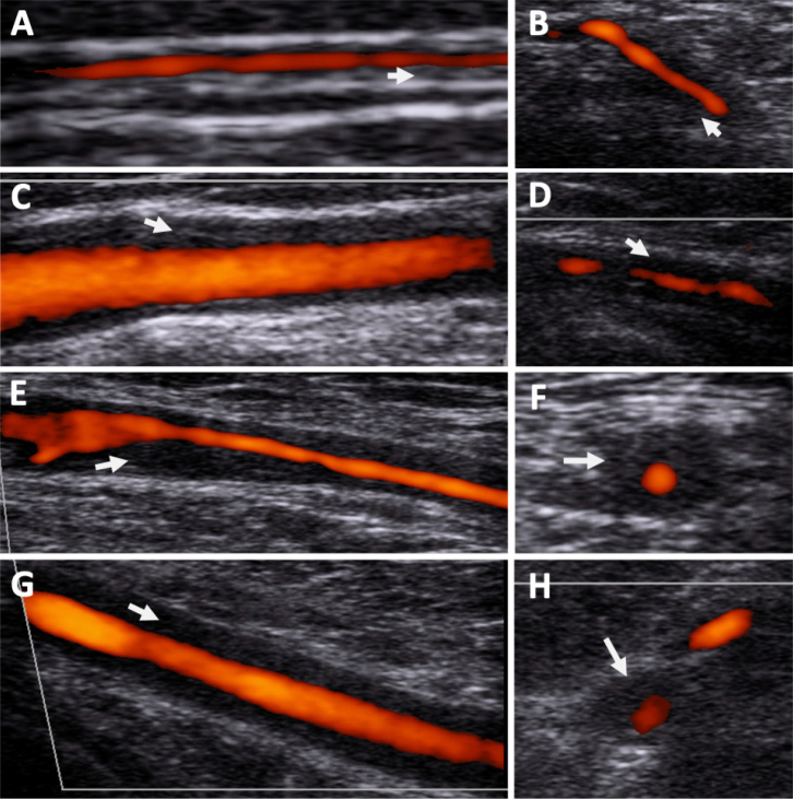
Figure 2.
Vascular ultrasound showing evidence of hypoechoic vessel wall thickening compatible with large vessel Vasculitis. (A) Right temporal artery parietal branch; (B) Left temporal artery common; (C) Right common carotid artery; (D) Left vertebral artery; (E) Right axillary artery; (F) Right axillary artery transverse plane; (G) Left subclavian artery; (H) Left subclavian artery transverse plane. Arrows show thickened vessel wall.

