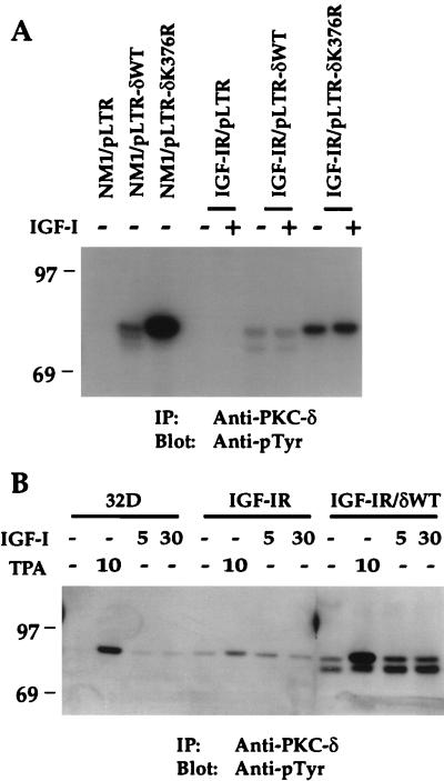
FIG. 6.
PKC-δWT and PKC-δK376R proteins are constitutively tyrosine phosphorylated in NM1- or IGF-IR-cotransfected NIH 3T3 cells. (A) NM1 and IGF-IR cotransfectants were serum starved overnight in DMEM and either untreated or stimulated with 10 ng of IGF-I per ml for 10 min. (B) 32D cells and transfectants were serum starved for 2 h and either untreated or stimulated with 100 ng of TPA per ml for 10 min or with 10 ng of IGF-I per ml for either 5 or 30 min. Equivalent cell lysates were immunoprecipitated with anti-PKC-δ serum. Transferred proteins were immunoblotted with anti-pTyr. Marker proteins are indicated in kilodaltons. IP, immunoprecipitation.

