FIG. 8.
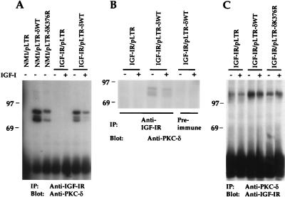
PKC-δWT and PKC-δK376R are constitutively associated with NM1 and IGF-IR in vivo. (A) Various NIH 3T3 transfectants were serum starved overnight in DMEM and either untreated or stimulated with 10 ng of IGF-I per ml for 10 min. Equivalent cell lysates were immunoprecipitated with anti-IGF-IR serum. Transferred proteins were immunoblotted with anti-PKC-δ. (B) Cell lysates were immunoprecipitated either with anti-IGF-IR or with preimmune serum. Transferred proteins were immunoblotted (Blot) with anti-PKC-δ. (C) The same lysates from panel A were immunoprecipitated with anti-PKC-δ serum. Transferred proteins were immunoblotted with anti-IGF-IR. Marker proteins are indicated in kilodaltons. IP, immunoprecipitation.

