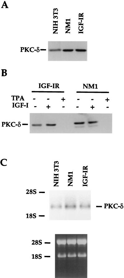
FIG. 9.
Endogenous PKC-δ protein and RNA are up-regulated by long-term IGF-IR activation. (A) NIH 3T3 cells and transfectants were maintained in media containing 10% calf serum and lysed. Equivalent amounts of proteins were loaded on SDS-PAGE gels. The transferred proteins were immunoblotted with anti-PKC-δ serum. (B) NIH 3T3 transfectants were serum starved for the first 8 h and either untreated or exposed to IGF-I (50 ng/ml) or TPA (100 ng/ml) for another 16 h. The cells were lysed, and transferred proteins from SDS-PAGE gels were immunoblotted with anti-PKC-δ. (C) Fifteen micrograms of total RNA from the parental NIH 3T3 and transfectants was isolated from normal cultured cells and loaded into agarose gel. Equivalent amounts of loading were demonstrated by ethidium bromide staining, as shown in the lower panel. The specific PKC-δ messages were detected with the full-length mouse PKC-δ as a probe (top panel). 18S and 28S rRNAs were used as markers.

