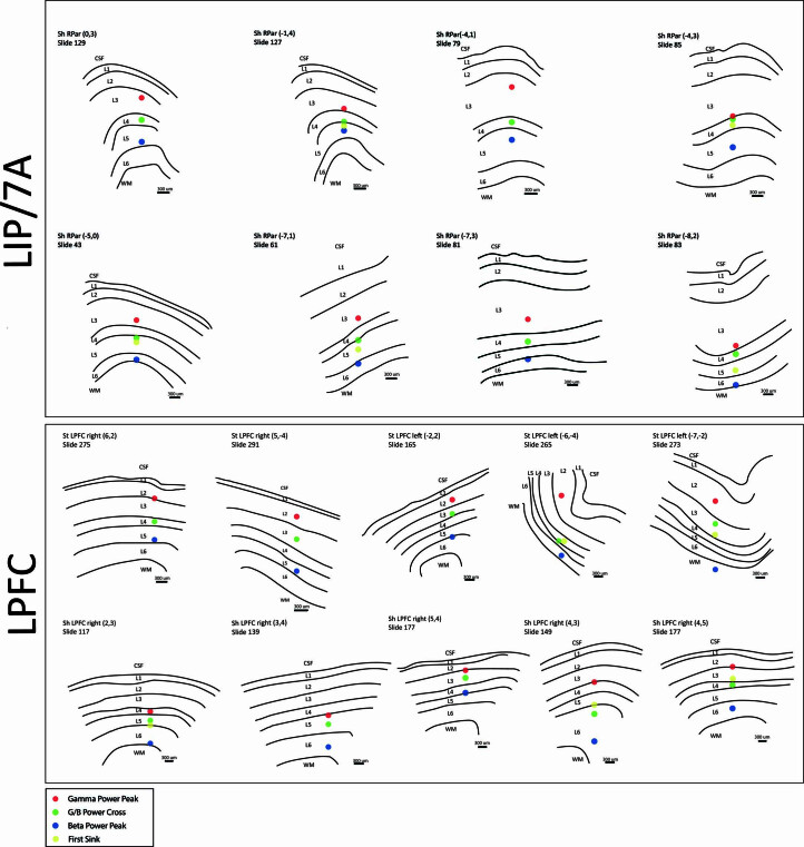
Extended Data Fig. 3. Individual anatomical probe reconstructions for parietal (LIP/7 A) and lateral prefrontal cortex.
Upper panel, traces of individual brain slices are shown for areas LIP/7 A with anatomically-defined layers labelled from cerebrospinal fluid (CSF), layer 1–6 (L1–L6), and white matter (WM). Each example includes monkey name, brain region, probe grid location, and histological slice number. Red, green, blue, and yellow dots correspond to gamma peak, relative power cross-over, alpha-beta peak, and CSD early sink respectively. Lower panel, traces of individual brain slices in LPFC.

