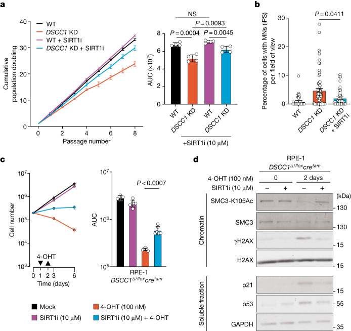
Fig. 4. SIRT1 inhibition rescues DSCC1-associated cellular phenotypes.
a, SIRT1i rescues the proliferation defect of DSCC1-mutant cells and decreases MN formation and accumulation. The proliferation of human iPS cells in which DSCC1 was disrupted (DSCC1 KD) using CRISPR–Cas9 (Extended Data Fig. 7) was compared with control cells (WT; parental line) as well as cells treated with SIRT1i. Statistical analysis was performed using two-tailed Student’s t-tests. n = 4 biological replicates. Data are mean ± s.e.m. b, SIRT1i (10 µM) treatment rescues MN formation in DSCC1-KD cells. Each dot represents an independent field of view. Data are mean ± s.e.m. Three biological replicates were performed. Significance was assessed by comparing the means of these experiments using a two-way Mann–Whitney U-test. c, Proliferation assay (left) and AUC (right) of the RPE-1 DSCC1Δ/floxcretam cell line in the presence of SIRT1i (10 µM) after DSCC1 deletion by 4-OHT treatment (addition and removal indicated by arrows). Data are mean ± s.e.m. Statistical analysis was performed using two-tailed Student’s t-tests, comparing the AUC for cells with and without SIRTi (10 µM) treatment. The experiment was performed three independent times (biological replicates) in duplicate. Significance was assessed by comparing the means of these experiments. d, Representative western blot images (from three independent/biological replicate experiments) showing chromatin fractionation of the RPE-1 DSCC1Δ/floxcretam cell line after the indicated treatments (uncropped images are shown in Supplementary Fig. 2).

