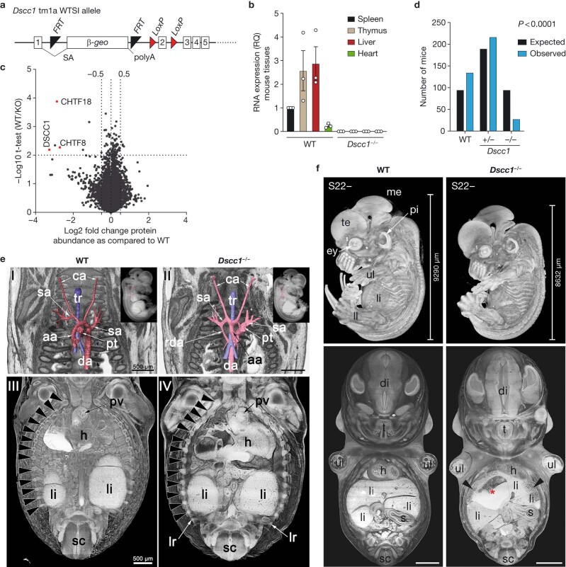
Extended Data Fig. 4. Dscc1 mutant mice show cardiac and vascular anomalies and reduced viability.
a, Diagram shows the targeting of the Dscc1 locus on mouse chromosome 15. A beta-galactosidase gene-trap including a splice acceptor site (SA) and a polyadenylation sequence (polyA) were inserted in intron 1 of the Dscc1 gene. Further elements were inserted to allow the generation of a conditional allele, such as FRT and LoxP sites. b, Bar graph showing quantitative PCR analysis of Dscc1 transcripts in adult mouse tissues. n = 3 mice with n = 5 technical replicates each. Mean is plotted with error bars representing s.e.m. c, Mass-spectrometry analysis of E13.5 embryo heads showing depletion of DSCC1, CHTF8 and CHTF18 proteins (members of the DSCC1-CHTF18-CHTF8 protein complex). The raw files were processed with Proteome Discoverer 2.4 (ThermoFisher) using the Sequest HT search engine and the analysis is presented in Source Data. Proteins/peptides were validated using Percolator. Only unique peptides were used for quantification. Red dots denote key significantly differentially expressed proteins (Student’s two-tailed t-test was used to determine significance). Two embryos of each genotype were analysed in this way. d, Mice born from Dscc1 heterozygous (+/−) intercrosses that survived past post-natal day 10 (P10) were genotyped and a Chi-squared analysis (two-tailed) was performed using the expected versus observed numbers of each genotype. Approximately a third of the expected Dscc1−/− mice survived past P10. e, Skeletal and vascular abnormalities in Dscc1−/− (right panels) embryos and for comparison control (left panels) embryos. Great intrathoracic arteries at developmental stage S22- (upper panels) are shown. Abnormal persistence of right dorsal aorta (rda) in a Dscc1−/− embryo. Surface models of the arteries in front of a coronal section through HREM data from anterior. Inlay shows the surface models inside a semitransparent volume model from right. Coronally sectioned semi-transparent volume models of thorax and abdomen from ventral (lower panels). The regular 13 ribs are indicated with arrowheads. Note the lumbar rib (lr) in the Dscc1−/− embryo. f, Growth delay and liver abnormalities in Dscc1−/− embryos. Control/wild-type (left panels) and Dscc1−/− (right panels) embryos are shown. Upper row: Growth and developmental delay can be seen in a E14.5 Dscc1−/− embryo relative to WT embryo. In addition, the developmental stage (S22-) of Dscc1−/− mutants is earlier than of wild-type littermates and as expected from reference data75. Lower row: Abnormal liver. Coronally sectioned semi-transparent volume models of thorax and abdomen from ventral. Blood filled cyst (red asterisk) and enlarged liver sinusoids (arrowheads). te, telencephalon; me, mesencephalon; ey, eye; pi, pinna; ul, upper limb; ll, lower limb; li, liver; tr, trachea; ca, common carotid artery; h, heart; pv, pulmonary valve; sa, subclavian artery; aa, ascending aorta; da, descending aorta; pt, pulmonary trunk; rda, right descending aorta, di, diencephalon; t, tongue; s, spleen; sc, spinal cord. For this experiment, n = 3 embryos/genotype were analysed.

