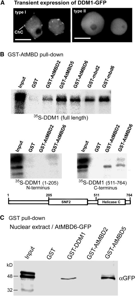
Figure 4.
AtMBDs Colocalize and Interact in Vitro with DDM1.
(A) DDM1 fused to GFP displays two types of subnuclear localization in Arabidopsis; type I, showing localization to chromocenters; type II, showing dispersion throughout the nucleus.
(B) GST-AtMBD proteins bind DDM1. GST pull-down assay was performed with the indicated AtMBD proteins fused to GST using in vitro–translated, 35S-labeled, full-length DDM1, N-terminal DDM1/1-205, or C-terminal DDM1/511-764. GST alone was used as a negative control. A schematic representation of the DDM1 protein and its unique domains is shown. Input indicates 15% of the input 35S-labeled proteins.
(C) GST-DDM1 precipitates AtMBD6-GFP from nuclear extract. GST alone, GST-DDM1, GST-AtMBD2, and GST-AtMBD5 were mixed with nuclear extract derived from transgenic Arabidopsis expressing AtMBD6-GFP. Precipitated proteins were resolved by SDS-PAGE and immunoblotted using anti-GFP to detect AtMBD6-GFP. Input lane indicates 10% of the input proteins.

