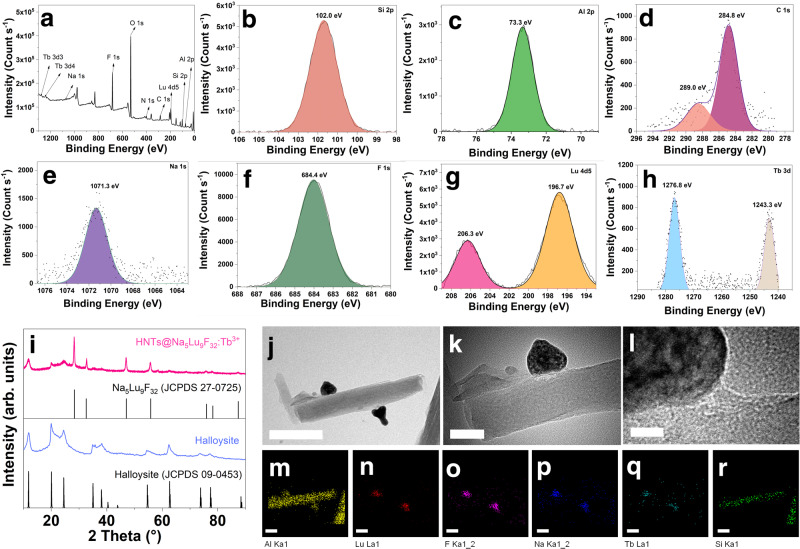
Fig. 2. Structural and micromorphology characterizations.
a X-ray photoelectron spectroscopy (XPS) pattern of Tb3+-doped Na5Lu9F32 anchored halloysite nanotubes (HNTs@Na5Lu9F32:Tb3+). b Si 2p region. c Al 2p region. d C 1 s region. e Na 1 s region. f F 1 s region. g Lu 4d5 region. h Tb 3d region. i X-ray powder diffraction (XRD) pattern of halloysite, HNTs@Na5Lu9F32:Tb3+, and standards. j Transmission electron microscopy (TEM) image of HNTs@Na5Lu9F32:Tb3+ (bar: 200 nm). k TEM image of HNTs@Na5Lu9F32:Tb3+ (bar: 50 nm). l High resolution TEM image of HNTs@Na5Lu9F32:Tb3+ (bar: 10 nm). m–r Elemental mapping analysis of HNTs@Na5Lu9F32:Tb3+ (m Al. n Lu. o F. p Na. q Tb. r Si; bar: 100 nm). Source data in a-i are provided as Source Data files.

