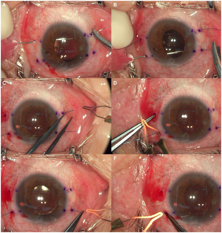
Figure 2.
(A) 7–0 polypropylene suture inserted through the temporal sclera, passed through the posterior surface of the iris plane, and externalized through nasal sclera. (B) The other suture inserted and externalized in the same manner. (C–F) Two temporal flanges were created via ophthalmic cautery and inserted into the scleral tunnel. Thereafter, two nasal flanges were created and then placed inro the scleral tunnels.

