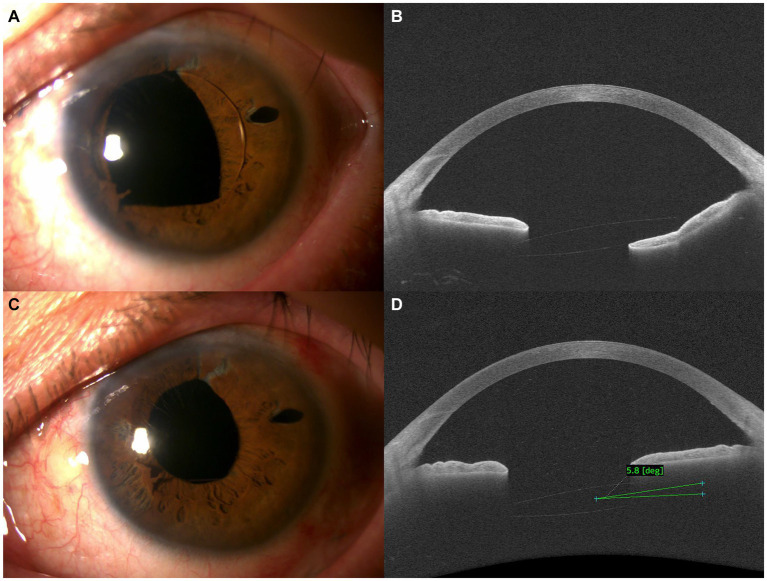
Figure 3.
(A,B) Preoperative anterior segment (A) and anterior segment optical coherence tomography (AS-OCT) images (B) demonstrating pupillary optic capture of the intraocular lens (IOL) after flanged IOL fixation. (C,D) Postoperative anterior segment (C) and AS-OCT images (D) demonstrating resolution of pupillary optic capture after IOL repositioning with a 7–0 polypropylene flange.

