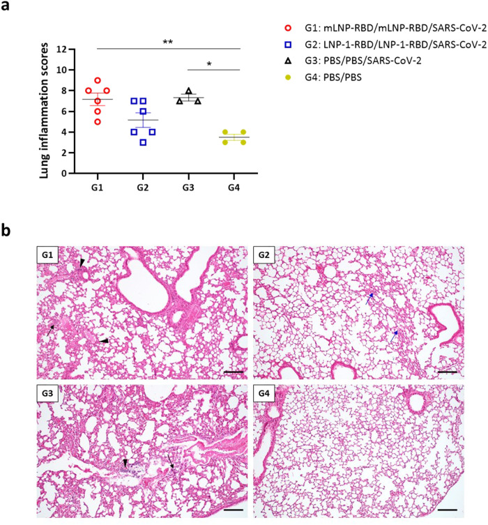
Fig. 5. Lung pathology in vaccinated and challenged transgenic K18-hACE2 mice.
a Lung inflammation scores observed in lung samples from vaccinated and challenged K18-hACE2 transgenic mice, and euthanized at day 7 (group 3) or day 14 post-challenge (groups 1, 2 and 4). Mean with SEM of cumulative histopathological lesion scores is indicated. An unpaired nonparametric Mann-Whitney test was performed.*p < 0.05; **p < 0.005. b Representative lung histopathological sections (H&E staining) from K18-hACE2 transgenic mice included in each of the experimental groups (scale bar: 200 µm). The severity and extent of inflammatory lung lesions observed in the immunized and challenged mice included in group 1 were similar to those described in the mice included in the non-immunized and challenged control group (group 3). Lesions included the presence of mild to moderate diffuse thickening of the alveolar septa, occasional small multifocal alveolar mononuclear cell infiltrates (black arrows) or mild multifocal perivascular and peribronchiolar mononuclear infiltrates (black arrowheads). Mice included in group 2 showed the lowest inflammatory scores. These animals showed only some lung areas with mild thickening of the alveolar septa (blue arrows) together with occasional small focal perivascular mononuclear infiltrates, showing an appearance highly similar to that observed in the PBS-treated non-challenged mice (group 4).

