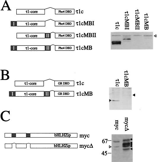
FIG. 4.
The myc boxes are signals for proteolysis. (A) Constructs used to investigate the effect of the myc boxes on protein levels in yeast cells (left). The τ1core (τ1c) of the human glucocorticoid receptor was fused to one or both of the myc boxes (indicated as shaded regions) and the Pho4 DBD. Whole-cell extracts from yeast strain YS33 expressing either the τ1c or the various τ1cMB constructs were analyzed by Western blotting (right) as described for Fig. 1A. The open arrowhead indicates a nonspecific band recognized by the antibody. (B) Schematic representation of the mammalian expression constructs used to investigate the destabilizing role of the myc boxes in COS7 cells (left). The myc boxes are shaded. COS7 cells were transiently transfected with the expression plasmids for τ1c and τ1cMB. A 30-μg portion of total protein from whole-cell extracts was subjected to SDS-PAGE (20% polyacrylamide) and blotted onto Hybond C-super membrane. The τ1c and τ1cMB proteins were detected with anti GR-DBD antibody (1:3,333 in 1% milk–phosphate-buffered saline–Tween) and secondary sheep anti-mouse IgG-HRP conjugate (1:2,000 in phosphate-buffered saline–Tween). Interaction with the antibodies was visualized by chemiluminescence (right). Solid arrowheads indicate the positions of the τ1c and τ1cMB proteins. (C) Western blot analysis of the levels of full-length c-myc proteins in mammalian cells. A schematic representation of the mammalian expression constructs used is shown on the left. The myc boxes are shaded. COS7 cells were transiently transfected with 0.1 μg of the expression plasmid pcDNA3myc or pcDNA3mycΔ. A 50-μg portion of total protein from whole-cell extracts subjected to SDS-PAGE (7.5% polyacrylamide) and blotted onto Hybond C-super membrane. The c-myc proteins were detected with anti-V5 antibody (1:10,000 in 1% milk–phosphate-buffered saline–Tween) and secondary sheep anti-mouse IgG-HRP conjugate (1:2,000 in phosphate-buffered saline–Tween). Interaction with the antibodies was visualized by chemiluminescence. The mycΔ protein is indicated by the solid arrowhead; the derivatives are indicated by solid circles. A nonspecific band is indicated by an open arrowhead. Standard molecular mass markers are indicated in kilodaltons.

