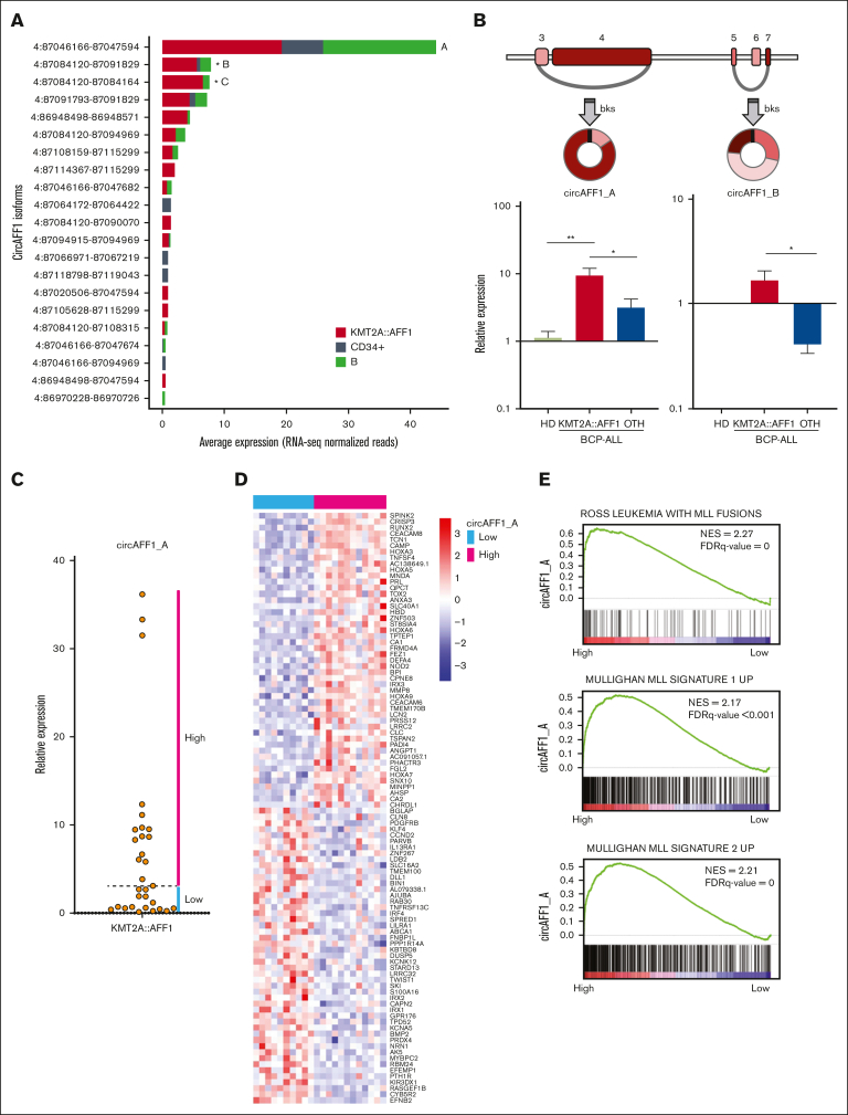
Figure 4.
CircAFF1_A and circAFF1_B isoform structure and expression in the extended patient cohort. (A) Expression levels of the circAFF1 isoforms in samples from patients with KMT2A::AFF1 ALL and CD34+ cells and B cells populations from HDs, according to RNA-seq analysis (significant DE is indicated by ∗). (B) Schematic representation of circAFF1_A and circAFF1_B structure (exons number according to ENST00000395146 transcript; colored boxes represent exons; gray segments, introns; and arches, circRNAs;) and bar graphs of expression levels quantified by qRT-PCR in bone marrow samples from HDs (N = 3), in pediatric patients with BCP-ALL, 31 with (KMT2A::AFF1, N = 31 for circAFF1_A and N = 17 for circAFF1_B) and 13 without (OTH) KMT2A::AFF1 rearrangement (mean ± standard error of the mean is shown; Mann-Whitney U test P value ∗P < .05 and ∗∗P < .01). (C) circAFF1_A level stratify patients into high- and low-expressors groups. (D) Patients with high and low circAFF1_A expression level are characterized by different genes expression profiles according to microarrays analysis (the top 50 ranked genes differentially expressed are shown in the heat map); (E) MLL signatures resulted positively enriched among genes differentially expressed between patients with high and low circAFF1_A expression. bks, backsplice site; FDR, false discovery rate.

