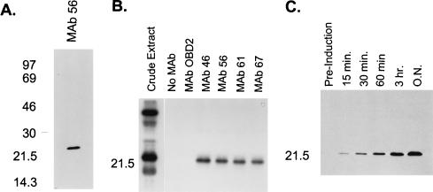
FIG. 2.
Identification of target protein. (A) Western blot analysis of crude mitochondrial lysate separated by SDS-PAGE on a 12% (wt/vol) gel. MAb 56 (and five other MAbs not shown) recognized an ∼21-kDa protein. (B) Immunoprecipitation of gRNA UV-cross-linked proteins. Proteins that cross-link with uniformly labeled gRNA in crude mitochondrial lysate and immunoprecipitate with the MAbs specific to gBP21 after treatment with RNase A were separated by SDS-PAGE on a 12% (wt/vol) gel and visualized by autoradiography as described in Materials and Methods. The four MAbs shown (and two others not shown) recognize an ∼21-kDa UV-cross-linking protein. (C) Western blot analysis showing that the MAbs (MAb 56 shown) recognize recombinant gBP21. Time points refer to sampling intervals after induction of culture (see Materials and Methods). Sizes are indicated in kilodaltons at the left.

