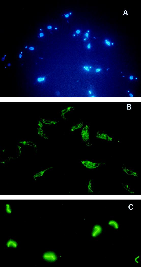
FIG. 3.
Immunofluorescence with MAbs specific to gBP21. Procyclic and bloodstream forms of T. brucei were fixed and stained with MAbs against gBP21 and with DAPI (see Materials and Methods). (A) DAPI staining showing the nucleus and smaller kinetoplast of procyclic T. brucei. (B) Procyclic T. brucei after incubation with MAb 56 and development with a goat anti-mouse antibody conjugated with fluorescein isothiocyanate. The kinetoplast is evident as an intensely staining spot in the network of mitochondrial tubules. (C) Bloodstream T. brucei examined as in panel B, showing the single tubular mitochondrion.

