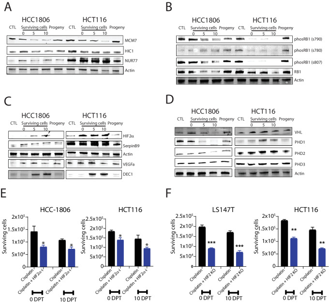
FIGURE 4.
Protein changes validate the role of HIF2α and RB1 for cell survival. A, Protein level changes of HIF2α-interacting proteins, MCM7, HIC7, and NUR77 in HCC1806 and HCT116 cells when untreated (CTL), when surviving at 0 DPT, 5 DPT, and 10 DPT and as progeny; demonstrated by Western blot analysis. Actin was used as a loading control. Molecular weight markers in kDa are shown to the left. B, Representative images of protein level changes of RB1 and its phosphorylated sites (s790, s780, and s807) in HCC1806 and HCT116 cells when untreated (CTL), when surviving at 0 DPT, 5 DPT, 10 DPT and as progeny; as determined by Western blot analysis. C, Protein level changes of HIF2α and its targets SERPINB9, VEGF, and DEC1 in HCC1806 and HCT116 cells when untreated (CTL), as surviving at 0 DPT, 5 DPT, and 10 DPT and as progeny; as determined with Western blot analysis. D, Protein level changes of VHL and PHD1–3 in HCC1806 and HCT116 cells when untreated (CTL), when surviving at 0 DPT, 5 DPT, and 10 DPT, and as progeny; as determined with Western blot analysis. E, Number of HCC1806 and HCT116 cells surviving at 0 DPT and 10 DPT when treated with cisplatin only or cisplatin together with the HIF2α inhibitor Belzutifan. F, Number of LS174T and HCT116 colon cancer cells surviving cisplatin at 0 DPT and 10 DPT as “normal” and with k HIF2α KO from biological replicates (n = 3) and P-value (∗∗, P < 0.01; ∗, P < 0.05; significant relative to vehicle) by ANOVA test as indicated.

