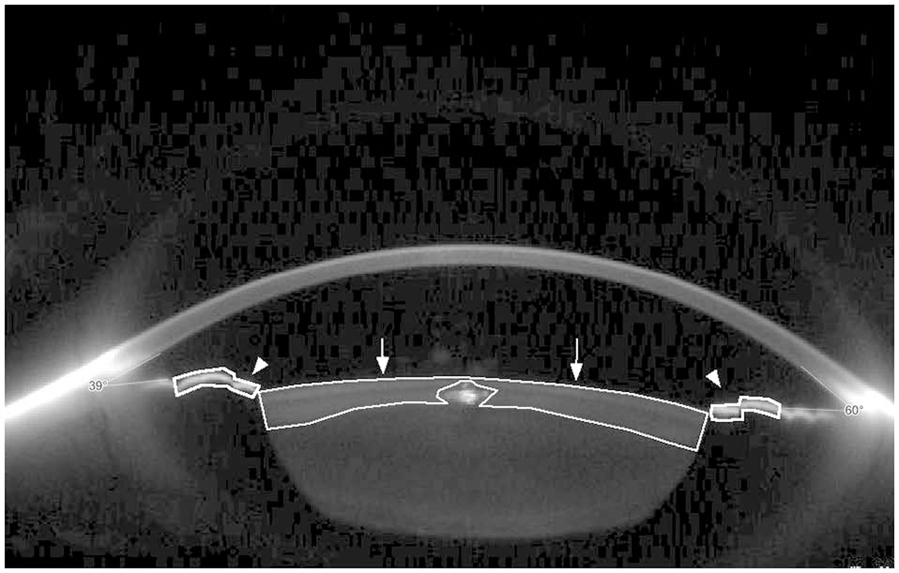
Figure 1.

Scheimpflug image mounted with ImageJ software to measure maximum density of anterior cortical lens and anterior iris surface. Selected anterior cortical lens (arrows) and iris anterior surfaces (arrowheads). Note that the intra-lenticular light artefact is excluded from the measurement.
