FIG. 6.
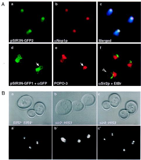
(A) SIR3N-GFP localizes to the nucleolus. The haploid strain UCC3107 was transformed with either a 2μ-based plasmid (a, b, and c) or a centromeric plasmid (d and f) expressing Sir3N fused to the green fluorescent protein under the control of the ADH promoter (pSIR3N-GFP2 and pSIR3N-GFP1, respectively). The direct fluorescence of Sir3N-GFP (green) (a), anti-Nop1p staining on the same cells visualized with a Cy3-conjugated secondary antibody (red) (b), and the merge of the two stainings (c) are shown. The blue area represents the nucleus. Colocalization of Sir3N-GFP and Nop1p is white. Cells transformed with pSIR3N-GFP1 and stained with anti-GFP antibodies (the kind gift of K. E. Sawin, Imperial Cancer Research Fund, London, England) visualized by a DTAF-conjugated secondary antibody (green) (d); the DNA staining of the same cells with POPO-3, which preferentially stains the nucleolar domain (red) (e); and anti-Sir2p immunofluorescence on a fixed wild-type diploid strain (GA229) that had been washed in 1% Triton–0.02% sodium dodecyl sulfate as described previously (10) (f) are also shown. Sir2p staining is visualized by a DTAF-conjugated secondary antibody (green), and the DNA is counterstained with ethidium bromide. Immunofluorescence assays were performed with affinity-purified antibodies as described in Materials and Methods. The arrows indicate an apparent looped body, while the arrowhead indicates the same loop extended. (B) SIR2 but not SIR4 is necessary for the enrichment of Sir3N-GFP in the nucleolus. The haploid strains UCC3107 (SIR+) (a and a′), UCC3203 (sir2::HIS3) (b and b′), and UCC3207 (sir4::HIS3) (c and c′) were transformed with plasmid pSIR3N-GFP1. The phase-contrast image (a to c) and the direct fluorescence (a′, b′, and c′) of Sir3N-GFP are shown. Results identical to those shown in c and c′ were obtained for a sir3::HIS3 strain.

