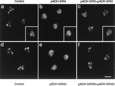
FIG. 9.
Overexpression of Sir3N restores the focal staining pattern of Sir3p. Yeast cells were stained with anti-Sir3p antibodies detected by a DTAF-conjugated secondary antibody (white signal). All signals are within the yeast nuclei, as indicated in the insets, where the anti-Sir3p signals are superimposed on a DNA stain to reveal the nuclear shape (see Materials and Methods). Strain EG37 (26) transformed with the vectors pAAH5 and pRS316 (a), pC-ASir4 (26) and pAAH5 (b), and pC-ASir4 and p2μ-ASir3 (c) and strain AJL275-2AVIIL transformed with the vectors pAAH5 and p2HG (d), pADH-SIR4C (pFP340) and pAAH5 (e), and pADH-SIR4C (pFP340) and pADH-SIR3N (f) are shown.

