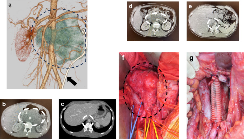
Fig. 1.
Computed tomography (CT) images and intraoperative findings. a The three-dimensional CT image captured before chemotherapy. The dotted line delineates the extent of the tumor. The black arrow indicates the ureteral stent. b, c CT images before chemotherapy. d, e CT images after chemotherapy. f Intraoperative findings. The dotted line indicates the extent of the tumor. g Intraoperative findings after tumor resection with abdominal aorta and reconstruction by prosthetic graft

