. 2024 Feb 14;20(2):229–231. doi: 10.3988/jcn.2023.0284

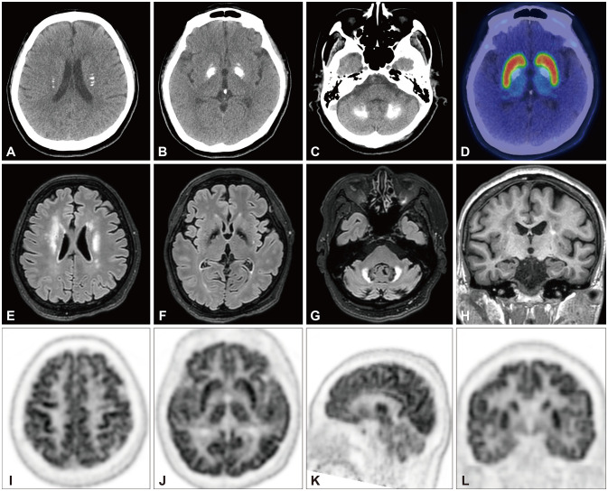
Fig. 1. Brain computed tomography showed dense calcifications in both basal ganglia, cerebellum, and corona radiata (A-C). 18F-FP-CIT PET showed preserved DAT uptake (D). Fluid-attenuated inversion-recovery MRI showed nonspecific hyperintensities in bilateral frontal white matter and the cerebellum (E-G). T1-weighted MRI showed mild mesial temporal lobe atrophy (H). Brain FDG PET showed decreased FDG uptakes in the frontal and parietal lobes and the cerebellum, while those in the sensorimotor cortex and basal ganglia were relatively preserved (I-L). 18F-FP-CIT, (3-[18F]fluoropropyl)-2β-carbon ethoxy-3β-(4-iodophenyl) nortropane; DAT, dopamine transporter; FDG, fluorodeoxyglucose; MRI, magnetic resonance imaging; PET, positron-emission tomography.

Fig. 1