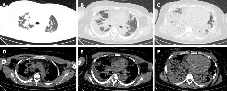
Figure 1.
Lung computed tomography examination in admission. There were diffuse multiple patchy heightening shadows in both lungs, with a partial bronchial inflation sign. A: Both upper lungs in the lung window; B: Middle lobe and tongue lobe in the lung window; C: Both lower lungs in the lung window; D: Both upper lungs in the mediastinal window; E: Middle lobe and tongue lobe in the mediastinal window; F: Both lower lungs in the mediastinal window.

