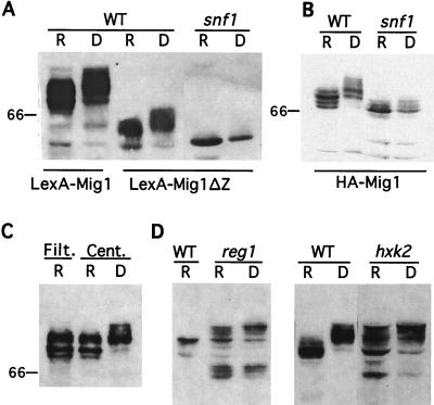
FIG. 2.
Immunoblot analysis of Mig1 fusion proteins. Cultures were grown in selective SC medium plus 5% glucose (repressed) (lanes R). Mid-log-phase cultures were derepressed by a shift to SC medium plus 0.05% glucose for 1 h (lanes D). Extracts were prepared, and proteins were separated by SDS-PAGE in 7.5% polyacrylamide and subjected to immunoblot analysis with anti-LexA (A, C, and D) or anti-HA (B). (A and B) Protein extracts (25 and 50 μg for wild-type [WT] and snf1-K84R strains, respectively) were prepared from strains MCY829 and MCY2692 transformed by pLexA-Mig1 or pLexA-Mig1ΔZ (A) and pHA-Mig1 (B). (C) Protein extracts (25 μg) were prepared from strain YM4738 expressing LexA-Mig1 from vector pJH106. Cells were collected by rapid membrane filtration (Filt.) or by centrifugation for 2 min (Cent.) (see Materials and Methods). (D) Protein extracts (25 μg for the wild type and 50 μg for reg1 and hxk2 strains) were prepared from strains FY250, MCY829, MCY3278, and MCY3541 transformed with pLexA-Mig1. The position of the 66-kDa size marker is indicated.

