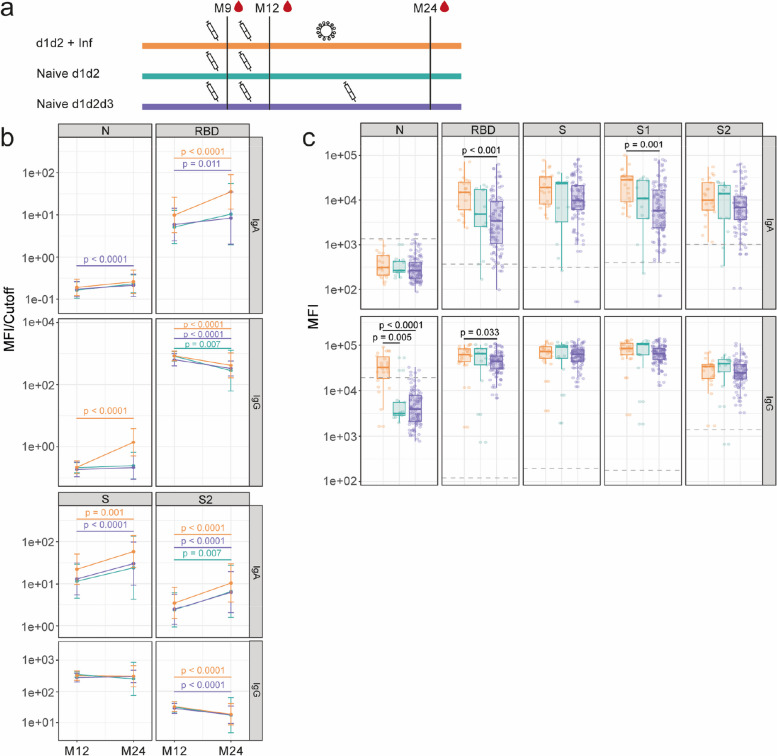
Fig. 2.
Evolution and comparison of antibody levels following hybrid immunity or vaccination alone. Plots show IgA and IgG levels against the receptor-binding domain (RBD) of the SARS-CoV-2 Spike glycoprotein (S), S, its subunits S1 and S2, and against the nucleocapsid (N). a Scheme illustrating the order and approximate time scale of vaccination and infection for each group. The syringe indicates a vaccine dose, the virus particle indicates an infection with SARS-CoV-2, and the blood droplet indicates plasma collection for antibody measurement. b Antibody levels (MFI/cutoff) at M12 and M24 time points in individuals vaccinated with 2 doses followed by an infection (n = 22), 2 doses without infection (n = 13), and 3 doses without infection (n = 129). The data shown are geometric mean in each group ± geometric SD. Statistical significance was tested by Wilcoxon signed-rank test and adjusted for multiple comparisons using Benjamini–Hochberg method. c Comparison of antibody levels (MFI) at M24 according to exposure. The center line on each box corresponds to the median MFI, the lower and upper hinges correspond to the first and third quartiles, and the whiskers extend from the hinge to the highest or lowest value within 1·5 × IQR of the respective hinge. Grey dashed line depicts the positivity cutoff for the given antibody. Statistical significance was tested by Wilcoxon rank-sum test and adjusted for multiple comparisons using Benjamini–Hochberg method. S1 antigen was only measured at M24

