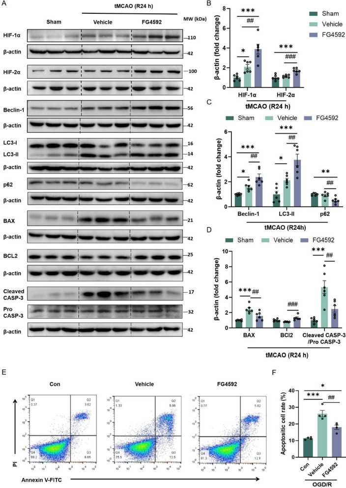
Fig. 2.
FG4592 activates autophagy and inhibits the apoptotic pathway after I/R injury. A Western blot images of HIF-1ɑ, HIF-2ɑ, Beclin-1, LC3-II, p62/SQSTM1, BAX, BCl2, cleaved-caspase-3 and caspase-3 in the peri-infarct brain tissues of Sham, vehicle and FG4592 groups after 24 hours of tMCAO. B-D Immunoblot analyses of proteins are shown in panel A. Data are presented as mean ± SEM. Sham vs vehicle or FG4592: ***p < 0.001, **p < 0.01, *p < 0.05. vehicle vs FG4592: ###p < 0.001, ##p < 0.01 (one-way ANOVA followed by Dunnett’s post-hoc test, n = 6 in each group). E and F Apoptotic cell analyses of HT-22 cells were detected using an Annexin V-FITC apoptosis flow cytometry. Cells underwent OGD for 3 hours and were treated with FG4592 or vehicle for 6 hours after reperfusion. The fractions of apoptotic cells were semi-quantified by Flowjo software. Data are presented as the mean ± SEM and from four independent experiments. Sham vs vehicle or FG4592: ***p < 0.001, *p < 0.05. vehicle vs FG4592: ##p < 0.01 (one-way ANOVA followed by Dunnett’s post-hoc test)

