Fig. 7.
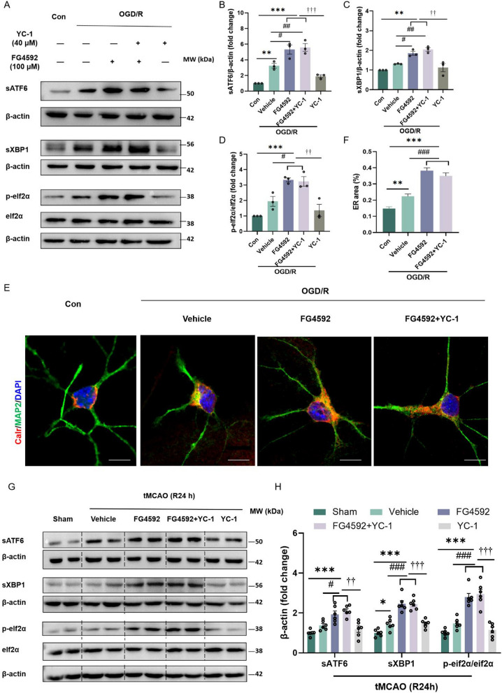
FG4592 also activates the UPR in a HIF-1α independent way in vitro and in vivo. A-D Immunoblots and quantitative analyses of sATF6, sXBP1 and p-eIF2ɑ in primary cortical neurons. Cells of YC-1 groups were pretreated with YC-1 for 1 hour previously. All experimental groups were treated with OGD for 3 hours and then treated with vehicle or FG4592 after reperfusion. Data are presented as the mean ± SEM, Con vs other groups: ***p < 0.001, **p < 0.01, vehicle vs FG4592: ##p < 0.01, #p < 0.05, FG4592 or FG4592+YC-1 vs YC-1: †††p < 0.001, ††p < 0.001 (one-way ANOVA followed by Dunnett’s post-hoc test, n = 3). E Representative confocal immunofluorescence images labelled with the ER marker calreticulin and the MAP2 in the primary cortical neurons. Scale bar = 10 μm. F The quantitative analysis of the ER area of different groups. Data are presented as mean ± SEM and were randomly from three independent coverslips. Con vs other groups: ***p<0.001, **p<0.01. Vehicle vs FG4592 or FG4592+YC-1: ###p < 0.001 (one-way ANOVA followed by Dunnett’s post-hoc test, n = 40 neurons in each group). G The representative western blots of sATF6, sXBP1 and p-eif2α in peri-infarct tissue of mice after 24 hours of tMCAO. Mice pretreated with YC-1 were injected with YC-1 intraperitoneally 1 hour before tMCAO. The experimental groups were treated with vehicle or FG4592 after tMCAO. H The statistical analysis of sATF6, sXBP1 and p-eif2α in the G panel. Data are presented as mean ± SEM. Sham vs other groups: ***p < 0.001, *p < 0.05. vehicle vs FG4592 or FG4592+YC-1 group: ###p < 0.001, #p < 0.05. YC-1 vs FG4592 or FG4592+YC-1 group: †††p < 0.001, ††p < 0.01 (one-way ANOVA followed by Dunnett’s post-hoc test, n = 6 in each group)

