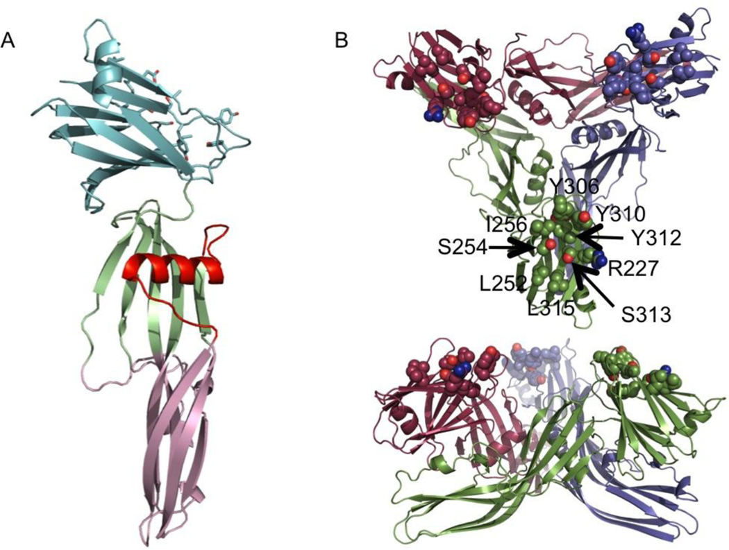
FIGURE 1.
Structure of full-length CPE. Cartoon representation of (A) the monomer, coloured pale green and pink for the oligomerisation domain and light cyan for the C-terminal claudin binding domain. The membrane-inserting residues are highlighted in red. (B) The trimer seen in the crystal. The monomers are coloured green, maroon and violet, the claudin-binding pockets are highlighted by drawing of likely claudin-binding residues as spheres.

