
Figure 2.
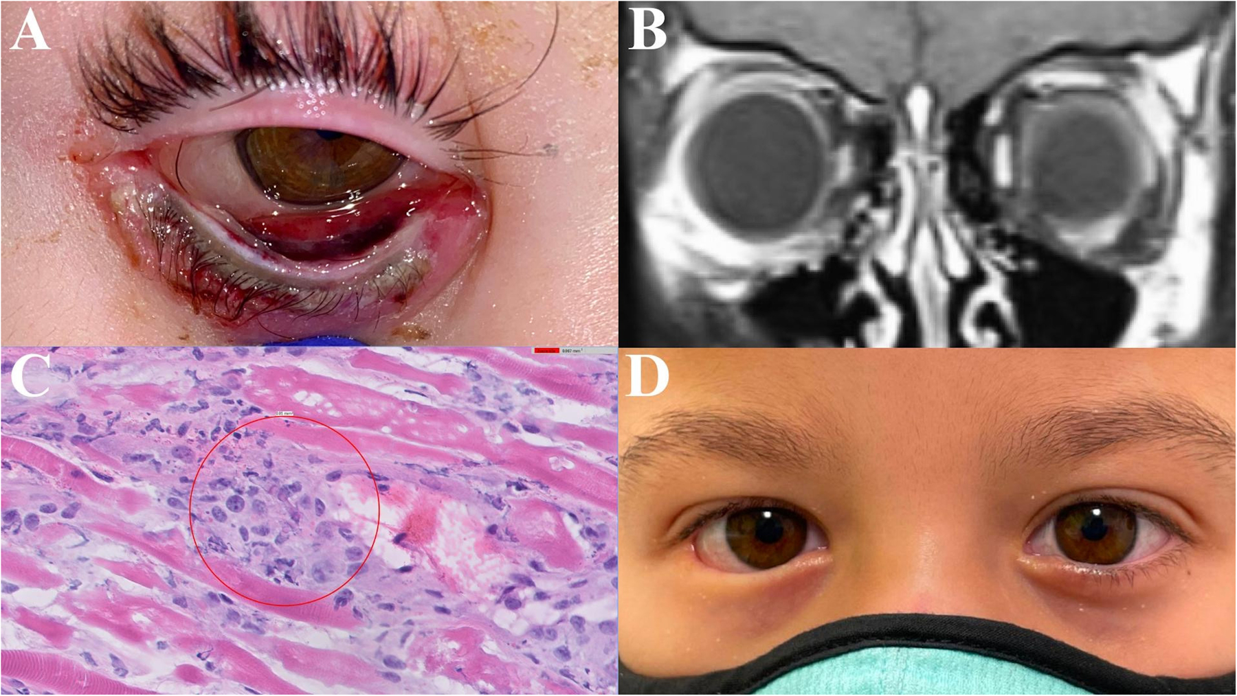
Case 2. A 4-year-old female with acute lymphoblastic leukemia (ALL) presenting with cutaneous mucormycosis of the right lower eyelid from disseminated pulmonary disease. A – Right lower eyelid edema, erythema, duskiness of the eyelid margin, and necrosis of the inferior palpebral conjunctiva. B – Magnetic resonance imaging (MRI) demonstrating enhancement of the right preseptal tissue. C – Right lower eyelid palpebral conjunctiva and pretarsal orbicularis histopathology demonstrating broad, ribbon-like, non-septate fungal hyphae (hematoxylin-eosin stain, original magnification 60x). D – Twenty months following treatment, the right lower eyelid maintains adequate protection of the ocular surface without further intervention.