
Fig. 2.
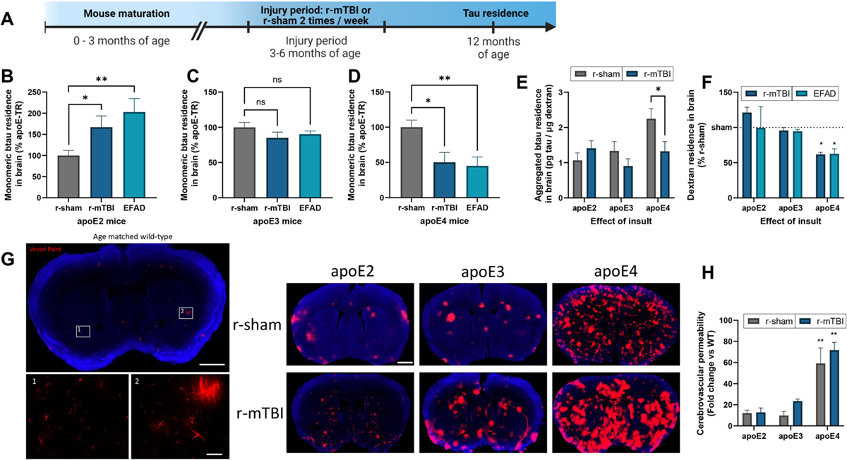
Influence of apoE and insult on tau brain residence.
(a) Timeline of closed head injury paradigm. (b-f) Following intracortical injection in 12 month old r-sham, r-mTBI and EFAD (apoE2, apoE3, and apoE4) mice, the amount of exogenous btau and co-injected LyD residing in the brain was determined at 2 h post injection. Monomeric (b-d) or aggregated (e) btau content was analyzed using an ELISA, while LyD (f) was analyzed via fluorescence. (b-d) Values represent mean + SEM (n = 5), and are expressed as the percentage tau residence normalized to each respective r-sham. *P < 0.05, **P < 0.01 as determined by one-way ANOVA and Bonferroni’s multiple comparisons test. (e) Values represent mean + SEM (n = 5) and are expressed as pg of aggregated btau per μg of LyD. *P < 0.05 as determined by one-way ANOVA and Bonferroni’s multiple comparisons test. (f) Values represent mean + SEM (n = 10) and are expressed as the percentage dextran residence normalized to each respective r-sham. *P < 0.05 as determined by two-way ANOVA and Bonferroni’s multiple comparisons test. (g, h) Vessel paint was applied to 12 month old r-sham and r-mTBI mice (apoE2, apoE3 and apoE4) via transcardiac injection. (g) Representative images of an age matched wild-type mouse showing vessel paint (red) leakage into the cortex with Dapi staining (blue). Insets 1 and 2 show boxed areas in (g). (h) Cerebrovascular permeability was quantified as the percent area of total cortex vessel paint signal normalized to age matched wild-type mice. Values represent mean + SEM (n = 3) and are expressed as the fold change normalized to age matched wild-type mice. *P < 0.05 as determined by two-way ANOVA and Bonferroni’s multiple comparisons test.