
Fig. 3.
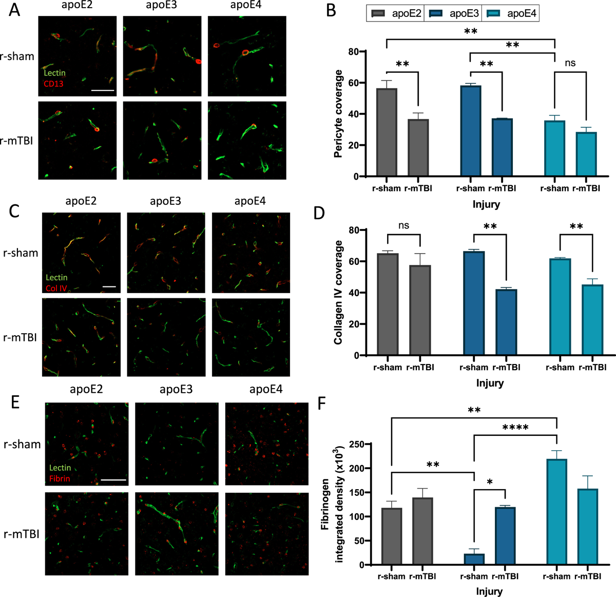
Influence of apoE and insult on pericyte coverage and BBB integrity.
(a) Confocal microscopy of CD13 immunodetection showing pericyte coverage (red) of lectin-positive brain capillaries (green) in the somatosensory cortex of 24 month old r-sham and r-mTBI mice (apoE2, apoE3 and apoE4). Scale bar, 25 μm. (b) Quantification of pericyte coverage on capillaries in r-sham and r-mTBI (apoE2, apoE3 and apoE4) mice. Values represent mean + SEM (n = 4). **P < 0.01 as determined by two-way ANOVA and Bonferroni’s multiple comparisons test. (c) Confocal microscopy of Collagen IV (red) and lectin-positive brain capillaries (green) in the somatosensory cortex of 24 month old r-sham and r-mTBI mice (apoE2, apoE3 and apoE4). Scale bar, 25 μm. (d) Quantification of Collagen IV coverage of lectin-positive capillaries. Values represent mean + SEM (n = 4). **P < 0.01 as determined by two-way ANOVA and Bonferroni’s multiple comparisons test. (e) Confocal microscopy of fibrin (red) and lectin-positive brain capillaries (green) in the somatosensory cortex of 24 month old r-sham and r-mTBI mice (apoE2, apoE3 and apoE4). Scale bar, 50 μm. (f) Quantification of extravascular pericapillary fibrinogen deposits. Values represent mean + SEM (n = 4). **P < 0.01 as determined by two-way ANOVA and Bonferroni’s multiple comparisons test.