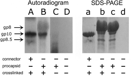
Figure 3.
Specific binding of [32P]pRNA I-i′ to connector demonstrated by UV crosslinking assay. Lanes A–D, 10% SDS–PAGE autoradiographed pictures; lanes a–d, the same gel stained by Coomassie blue. Lane A, pRNA crosslinked to connector and then treated by RNase A; lane B, procapsid crosslinked to pRNA and then treated by RNase A; lane C, procapsid–pRNA complex without crosslinking, and then treated by RNase A; lane D, pRNA treated by RNase A. Lanes a, b, c and d correspond to A, B, C and D, respectively.

