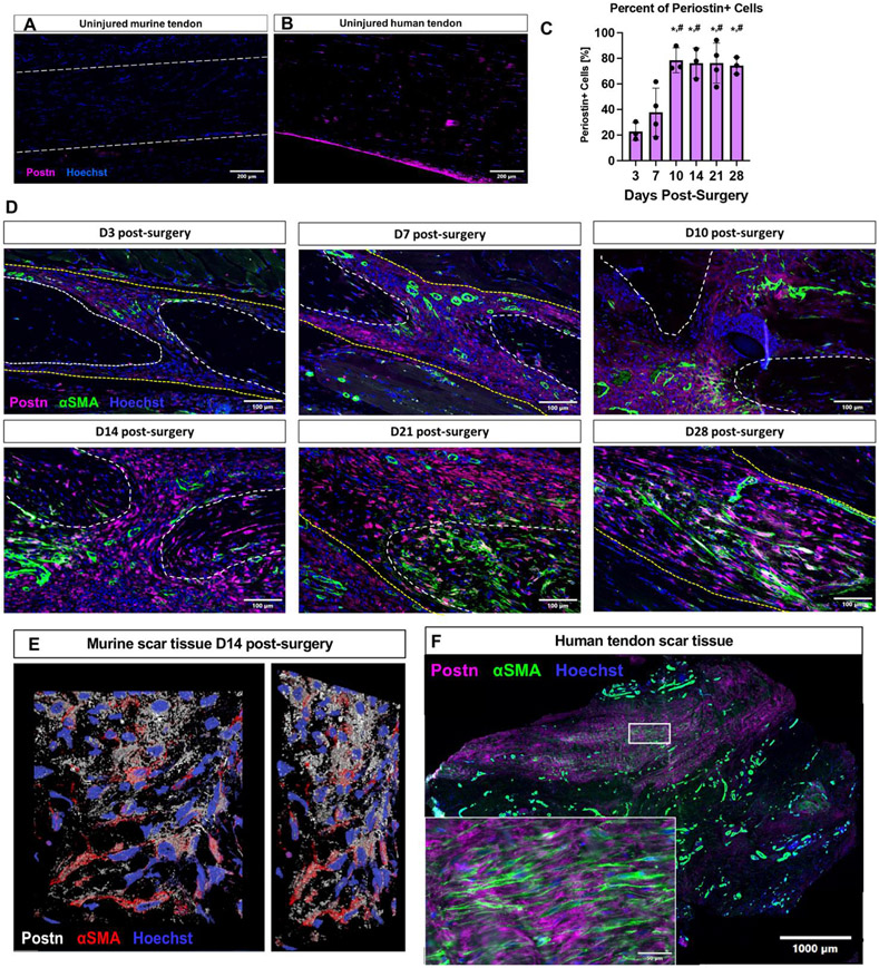
Figure 1. Periostin expression is dynamic during tendon healing.
(A) During homeostasis minimal Periostin (Postn, pink) expression is observed in C57Bl/6J murine flexor digitorum longus (FDL) tendons, while Postn expression was observed in the epitenon of uninjured human flexor tendon samples (B). (C & D) C57Bl/6J mice underwent FDL repair surgery and were harvested for histology at D3, 7, 10, 14, 21, and 28. Tissue sections were stained for αSMA (green), secreted Postn (pink), and Hoescht (nuclei, blue), the percent of Periostin+ cells was quantified over time. (E) A 2D representation of a 3D confocal z-stack imaged from an area of the murine scar tissue at D14 post-surgery. Postn staining is shown in white, αSMA in red, and nuclei in blue. (E) Human tenolysis tissue was processed for paraffin sectioning and stained for αSMA (green), Postn (pink), and Hoescht (nuclei, blue). Inset is magnified from the area enclosed by the box in the low power image. (*) indicates p<0.05 vs D3, (#) indicates p<0.05 vs D7.

