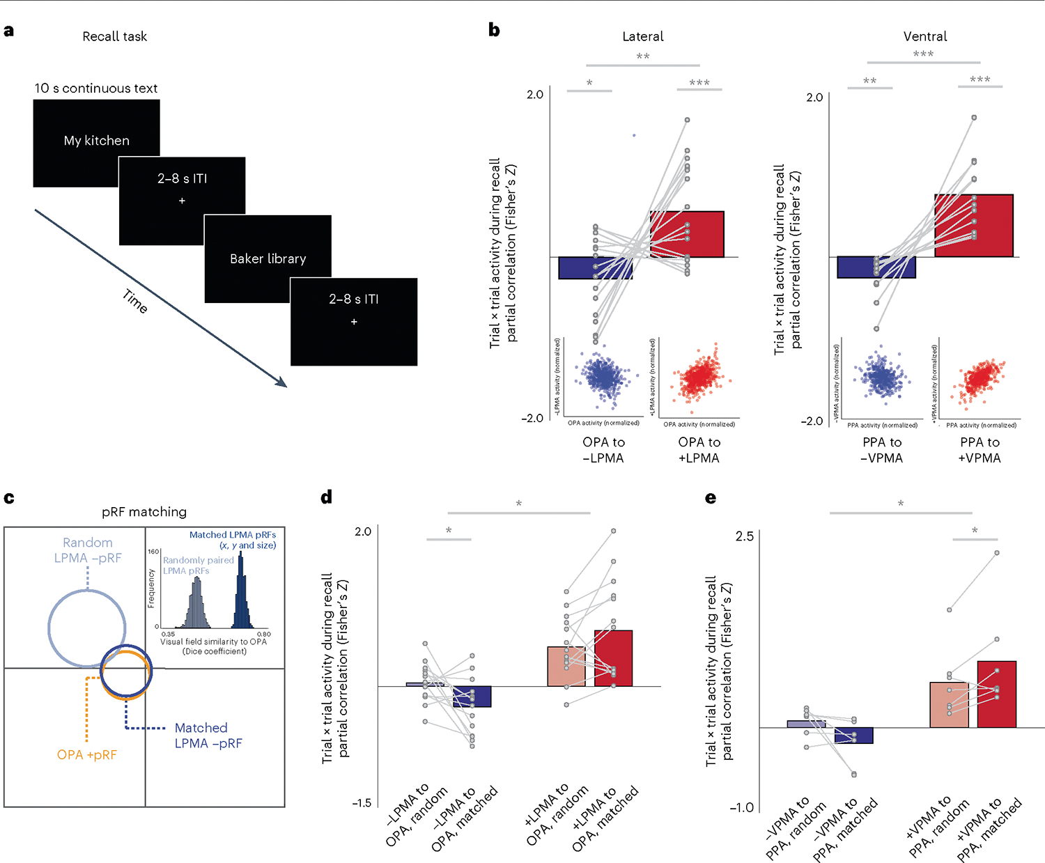
Fig. 5 |. Positive pRFs in SPAs and negative pRFs in PMAs exhibit a spatially specific push–pull dynamic during memory recall.

a, In Exp. 2, participants visually recalled personally familiar places for 10 s (cued with stimulus name; 36 trials per participant), and we calculated activation versus baseline at each trial. b, Negative and positive pRF (−pRF, +pRF) populations in SPAs and PMAs exhibit an opponent interaction during recall. Correlation in trial × trial activation of pRFs in SPAs was compared with +/−pRFs in PMAs using paired-sample t-tests. On both the lateral and ventral surfaces, when the activity of − pRFs in the PMA is high, activity in the corresponding SPA is reduced. By contrast, when activity in +pRFs in a PMA is high, activity in the adjacent SPA is also high. Inset, scatter plots show normalized trial-wise activation of each region for all trials in all participants. c, Schematic of pRF matching between OPA with −pRFs in LPMA. Schematic pRFs are depicted as circles. The OPA pRF is shown in orange, along with its best matched −pRF in LPMA (shown in dark blue) and randomly matched −pRF in LPMA (light blue). Inset, the histogram indicates the Dice coefficients for iterative matching between OPA pRFs and randomly paired pRFs from LPMA (light blue) compared to matched −pRFs from LPMA (dark blue) for one example participant, showing better correspondence between matched and random ‘unmatched’ pRFs. This pattern was consistent in all participants. d,e, Matched pRFs had a significantly stronger association in trial × trial activation than unmatched pRFs on the lateral (d) and ventral (e) surfaces, which suggests that the interaction between the perceptual and memory areas is spatially specific (paired t-tests) *Ptwo-tailed< 0.05, **Ptwo-tailed < 0.01, ***Ptwo-tailed < 0.001.
