FIG. 8.
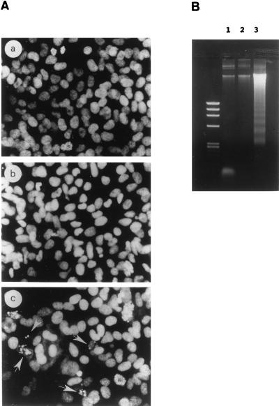
Apoptosis in RARγ1 antisense transgene-transfected SK-N-BE2(c) cells. (A) Morphological analysis of propidium iodide-stained nuclei from control cells (a) compared to RARγ1-overexpressing cells (b) and RARγ1 antisense transgene-transfected cells (c). Nuclei with typical morphological features of apoptosis are indicated (arrows). (B) Agarose gel electrophoresis of DNA from mock-transfected SK-N-BE2(c) cells (lane 1), RARγ1-overexpressing cells (lane 2), and RARγ1 antisense transgene-transfected cells (lane 3). Identical numbers of cells from each sample were lysed. DNA was isolated and electrophoresed on a 1.2% agarose gel. The left lane contains molecular size markers (φX174 RFDNA/HaeIII fragments [GIBCO]).

