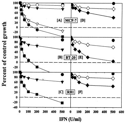
FIG. 1.
Cells were grown in the presence of various doses of IFNs and RA (1 μM) for 7 days. At the end of the experiment, the cells were fixed and stained with sulforhodamine B as described in Materials and Methods. The absorbance at 570 nm of bound dye was quantified and expressed as a percentage of untreated controls. Each datum point is the mean ± standard error (SE) of six replicates. Symbols: ○, IFN-α; ▿, RA plus IFN-α; ▾, IFN-β; ■, RA plus IFN-β; ◊, IFN-γ; ⧫, RA plus IFN-γ; •, RA. Human MCF-7 and BT-20 and murine RB1 breast carcinoma cells were treated with human and murine IFNs, respectively. Absorbance values for 0 and 100% growth in this assay, respectively, are as follows: MCF-7, 0.185 and 1.75; BT-20, 0.201 and 2.03; and RB1, 0.172 and 1.59. Values on the negative scale indicate death of initially plated cells.

