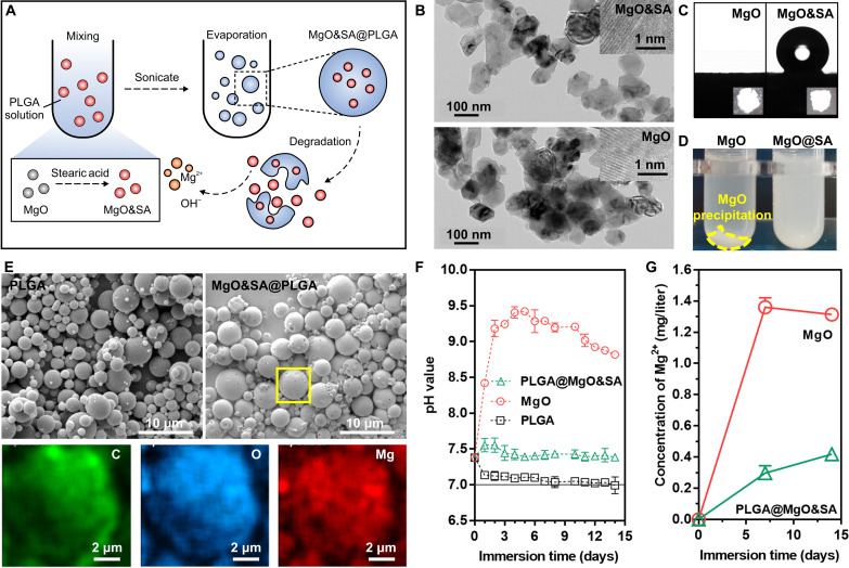
Fig. 6. Preparation and characterization of engineered MgO&SA@PLGA microspheres.
(A) Schematic diagram of the preparation of engineered MgO&SA@PLGA microspheres. (B) Transmission electron microscopy (TEM) images. (C) Water contact angle (CA). (D) Dispersibility of MgO nanoparticles after treatment with SA. (E) SEM images and EDS mapping (corresponding to the yellow box of MgO&SA@PLGA). (F) pH value change and (G) Mg2+ release in PBS at 37 ± 0.5°C as a function of time (14 days).

