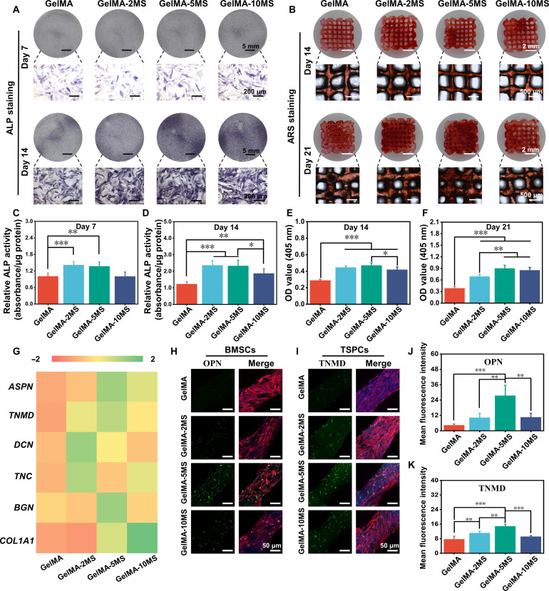
Fig. 3. The osteogenic differentiation of BMSCs and tenogenic differentiation of TSPCs encapsulated in the multicellular scaffolds.
(A) The ALP staining of multicellular scaffolds containing different concentrations of MS nanoparticles after being cultured for 7 and 14 days. (B) The alizarin red S (ARS) staining of multicellular scaffolds containing different concentrations of MS nanoparticles after being cultured for 14 and 21 days. The relative quantitative analysis of ALP staining after (C) 7 and (D) 14 days of culture and ARS staining after (E) 14 and (F) 21 days of culture (n = 5). (G) The tenogenic differentiation–related cytokines (ASPN, TNMD, DCN, TNC, BGN, and COL1A1) secretion of TSPCs in multicellular scaffolds with different concentrations of MS nanoparticles during the culture of 14 days (n = 3). The expression of (H) OPN protein in BMSCs and (I) TNMD protein in TSPCs within multicellular scaffolds containing different concentrations of MS nanoparticles. The corresponding semiquantitative analysis of (J) OPN and (K) TNMD protein expression (n = 3). *P < 0.05, **P < 0.01, and ***P < 0.001. MS nanoparticles held the significant ability to simultaneously induce tenogenic differentiation of TSPCs and osteogenic differentiation of BMSCs in the multicellular scaffolds.

