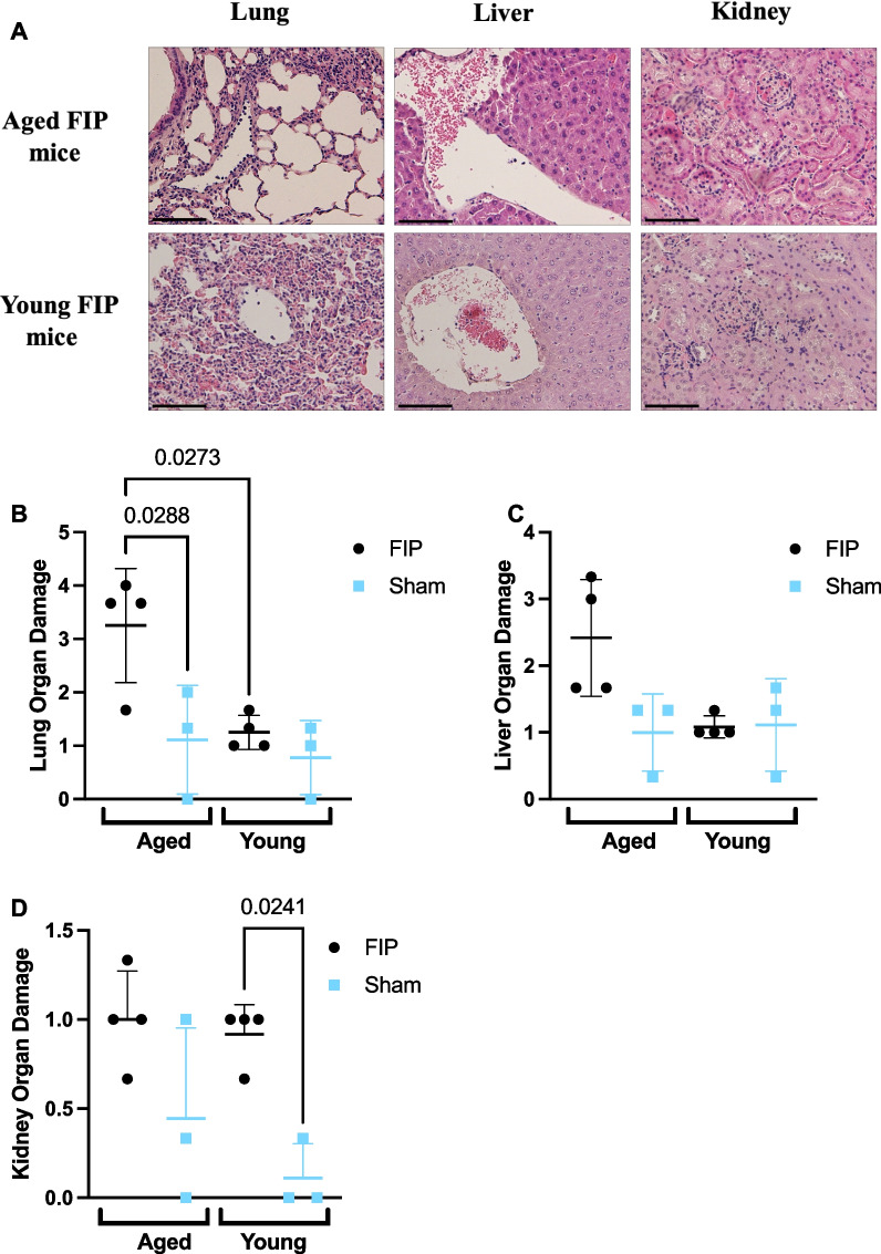
Fig. 3.
Histology scores in the lung, liver, and kidney in aged and young mice during the early phase of sepsis (12 h time course study). Representative images of the lung, liver, and kidney sections stained for H&E in aged and young mice at 8 h post-FIP (A). Organ injury scores for the lung (B), liver (C), and kidney sections (D) were based on inflammation, thrombosis, and organ morphology. Aged FIP (n = 4), young FIP (n = 4), aged sham (n = 3), and young sham (n = 3). Data are presented as mean ± SD and analyzed using a one-way ANOVA. P-values < 0.05 were considered significant. Scale bars represent 50 m

