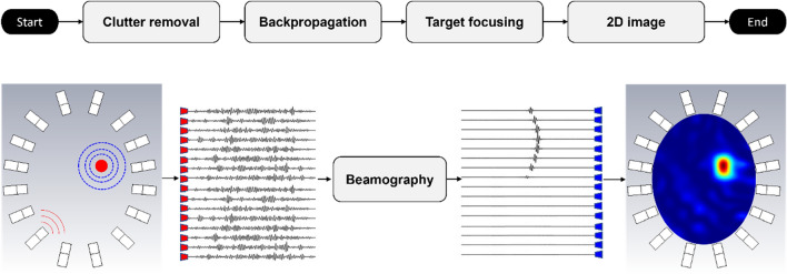
Figure 5.
Flowchart of beamography, and an example. Beamography detects and localizes strokes by performing two tasks (1) clutter mitigation, and (2) target focusing. Clutter mitigation is accomplished through symmetry and average subtractions. Marginal distortions due to the inevitable anatomical brain asymmetry and the head displacement are alleviated by average subtraction, where the average of all signals at all frequency samples is subtracted from each of them. After clutter removal, the data captured by each sensor is back-propagated to the imaging domain, using the domain Green’s function to calculate the field observed at each point in the brain for each transmitter–receiver pair and each frequency sample. Those calculated fields are then superposed to construct the final image. The location of the stroke in this method is shown by a higher intensity compared to healthy tissues.

