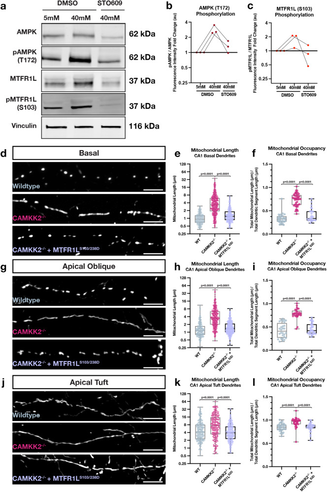
Fig. 9. Activity-dependent and Camkk2-dependent phosphorylation of MTFR1L by AMPK mediates compartmentalized mitochondria morphology in dendrites of CA1 PNs in vivo.
a Western blots of whole cell lysates from mouse hippocampal neurons maintained in culture for 18-21DIV and treated for 15 min with physiological (5 mM) extracelllular potassium chloride (KCl) (first column) or high KCl (40 mM) inducing membrane depolarization (columns 2 & 3) in the presence (column 3) or absence (columns 1 & 2) of the Camkk2 inhibitor STO609. These results demonstrate that phosphorylation of AMPKα catalytic subunit on T172 is increased by neuronal depolarization which is blocked by STO609. In turn, AMPK phosphorylation of its substrate MTFR1L on S10325 is increased by depolarization which is Camkk2-dependent since it is blocked by STO609. b Quantification of western blots with fold change of fluorescence intensity of pAMPK normalized to total AMPK plotted for each condition relative to 5 mM KCL treatment. c Quantification of western blots with fold change of fluorescence intensity of pMTFR1L normalized to total MTFR1L plotted for each condition relative to 5 mM KCL treatment. (d-l) Rescue experiments showing that phosphomimetic form of Mtfr1l (Mtfr1lS2D) mimicking phosphorylation by AMPK25 is sufficient to rescue compartmentalized mitochondria morphology in basal (d–f), apical oblique (g–i) and apical tufts (j–l) dendrites of CA1 PNs in vivo. CA1 PNs from wild-type (WT) or Camkk2-/- constitutive knockout mice were IUE with the same mitochondrial markers/cell fills as in Fig. 3 (WT and Camkk2-/-) and a plasmid cDNA expressing phosphomimetic mutant on the two serine residues phosphorylated by AMPK (S103D and S238D) of Mtfr1l (Mtfr1lS2D)25. Data and quantifications from WT and Camkk2-/- are the same as in Fig. 6. Basal Dendrites (WT and Camkk2-/-: See Fig. 6; Camkk2-/- + Mtfr1lS2D: n = 28 dendritic segments, 375 individual mitochondria, mean length = 1.583 μm ± 0.061 (SEM), mean occupancy = 39.99% ± 2.12%). Apical Oblique Dendrites (WT and Camkk2-/-: See Fig. 3; Camkk2-/- + Mtfr1lS2D: n = 37 dendritic segments, 459 individual mitochondria, mean length = 1.743 μm ± 0.053 (SEM), mean occupancy = 46.19% ± 2.22%). Apical Tuft Dendrites (WT and Camkk2-/-: See Fig. 3; Camkk2-/- + Mtfr1lS2D: n = 30 dendritic segments, 315 individual mitochondria, mean length = 5.310 μm ± 0.262 (SEM), mean occupancy = 84.00% ± 1.31%). Experiments in a were replicated 3 times. For (e), f, (h), (i), (k), and (l), p values are indicated in the figure following a one way ANOVA with Sidak’s multiple comparisons test. For (b) and (c), individual points from independent experiments are shown with connected lines. For (e), (f), (h), (i), (k), and (l), data are shown as individual points on box plots with 25th, 50th and 75th percentiles indicated with whiskers indicating min and max values. Scale bars, 5 μm.

