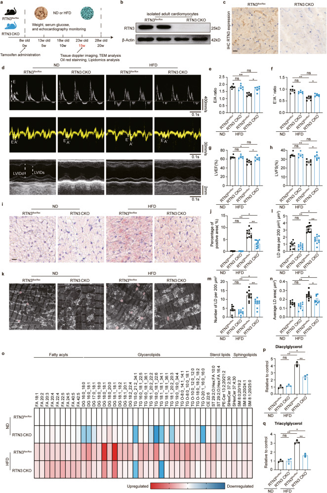
Fig. 3. Cardiomyocyte-specific RTN3 deletion ameliorated HFD-induced cardiac dysfunction and myocardial lipid accumulation.
a Diagram of the RTN3 knockout construction and high fat diet protocol; b Representative western blotting images of RTN3 protein expression in isolated adult cardiomyocytes; c Representative immunohistochemical images indicating RTN3 protein level, scale bar = 40 μm; d–h echocardiographic assessment was performed on RTN3 flox/flox and RTN3 CKO mice after 15-week ND or HFD. n = 6 mice each group. Representative doppler, tissue doppler, and M-mode echocardiography images (d) and quantitative analysis of E/A ratio (e), E′/A′ ratio (f), LVEF (g), and LVFS (h); i Representative oil red staining images indicating intramyocardial lipid content, scale bar = 20 μm; j Quantitative analysis of positive area of oil red staining (n = 10 images each group); k Representative TEM images of myocardium, LDs were labeled as red. Scale bar = 2 μm; Quantitative analysis of LD area per 200 μm2 (l), LD number per 200 μm2 (m), and average LD area (n) (n = 10 images each group); o Heatmap of metabolic alterations organized by lipid class (n = 3 mice each group); Quantitative analysis of diacylglycerol (p) and triacylglycerol (q) (n = 3 mice each group). Data are expressed as Mean ± SEM. Differences are significant for *P < 0.05, **P < 0.01.

