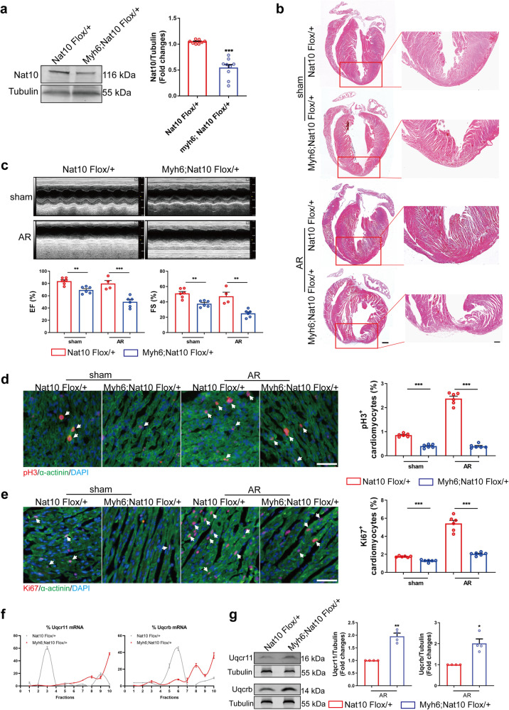
Fig. 6. The regenerative ability of cardiomyocyte-specific Nat10 knockdown mice.
a Representative western blots and statistic data showing Nat10 expression in the hearts from adult Nat10 knockdown mice (Myh6;Nat10 Flox/+) and control mice (Nat10 Flox/+) (Nat10 Flox/+, n = 8 mice; Myh6;Nat10 Flox/+, n = 9 mice). ***p < 0.001 vs. Nat10 Flox/+. b Morphology of Nat10 Flox/+ and Myh6;Nat10 Flox/+ mouse hearts at 28 days after sham or AR surgery analyzed by HE staining (n = 3 mice). Scale bar: 500 μm (left) and 200 μm (right). c Echocardiographic analysis of Nat10 Flox/+ and Myh6;Nat10 Flox/+ mice (n = 6, 6, 4, 6 mice per group). **p < 0.01, ***p < 0.001 as indicated. d Representative heart tissue sections stained with pH3 at P7 after AR, and the quantification of the percentage of pH3+ cardiomyocytes (n = 6 mice). ***p < 0.001 as indicated. White arrows point the positive cardiomyocytes. e Representative heart tissue sections stained for Ki67 at P7 after AR, and the quantification of the percentage of Ki67+ cardiomyocytes (n = 6 mice). ***p < 0.001 as indicated. White arrows point the positive cardiomyocytes. f Polysome profiling analysis of Uqcrb and Uqcr11 (n = 3 mice). g Western blot analysis of Uqcr11 and Uqcrb in Nat10 Flox/+ and Myh6;Nat10 Flox/+ mouse hearts (n = 4 mice). *p < 0.05, **p < 0.01 vs. Nat10 Flox/+. Two-tailed Student’s t test with Welch’s correction a, e, g. Two-tailed Student’s t test c–e. Data are presented as mean ± SEM. Scale bar: 50 μm d, e. Source data are provided as a Source Data file. Exact p values are provided in the Source Data file.

