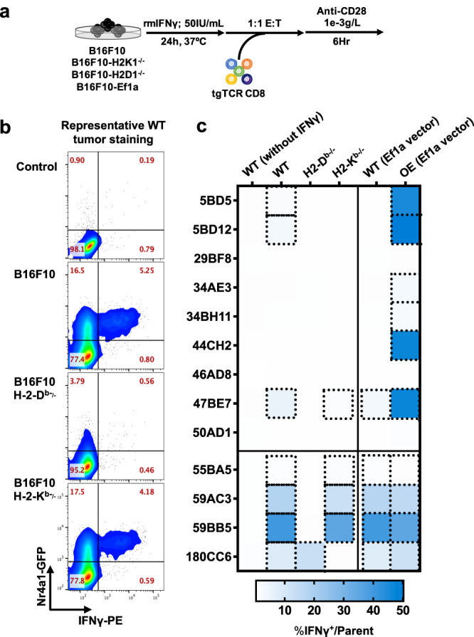
Fig. 3. Hsf2 neoantigen-reactive CD8+ T cells recognise tumour cells in vitro.

a Wild type B16F10 (WT), B16F10 lacking either MHC-I H2-Db or H2-Kb (B16F10-H2Db−/− and B16F10 H2Kb−/−, respectively) or B16F10-Ef1a (overexpressing neoantigenic or TAA peptide) were plated, exposed to recombinant murine IFNγ (rmIFNγ), and T cells expressing tgTCRs engineered from Nr4a1-eGFP mice were added at a 1:1 effector:target (E:T) ratio and co-incubated. Nr4a1-GFP is a marker of TCR signal transduction. b Cytokine production was measured by intracellular flow cytometry (n = 3 biological replicates/condition, repeated 3 times). Representative flow cytometric analysis of CD8+TCR-47BE7+ (Hsf2-reactive) cells exposed to (WT) B16F10 target cells is shown. Gating strategy for flow cytometry is shown in Supplementary Fig. 5. c Heatmap shown summarises the frequency of IFNγ+ cells of the parent CD8+tgTCR+ population. TCR clone names are shown for each row. IFNγ was withheld from conditions shown in the leftmost heatmap column (indicated as ‘WT (without IFNγ)’ to serve as a negative control. ‘WT (Ef1a vector)’ indicates that B16F10 was transduced with an “empty antigen” Ef1a lentiviral construct, meanwhile ‘OE (Ef1a vector)’ indicates that B16F10 was transduced (for antigen overexpression [OE]) with the appropriate antigen matching the TCR indicated.
