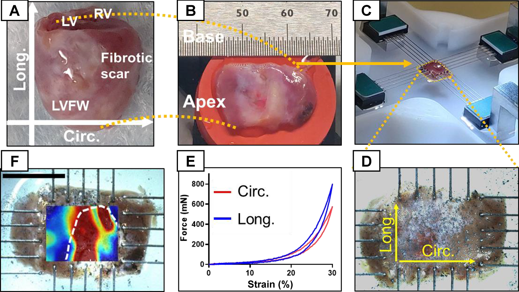
Fig. 2.

LVFW dissection and testing process. (A) Heart specimen with the atria removed. (B) Excised LVFW with apex and base labeled. (C) Mechanical testing of the LVFW specimens subjected to biaxial tensile loads using 5-rake tines per side. (D) Magnified image of LVFW testing with directions marked for reference. A dense, random, and multi-sized spread of graphite powder was used to obtain the regional strain field in LVFW specimens using DIC. (E) Representative force-strain relationship obtained from biaxial testing. (F) Representative strain contour obtained from DIC imaging of a 3-wk specimen. The white dashed line approximates the scar region identified from histology. Circ: circumferential direction; Long: longitudinal direction; LV: left ventricle; LVFW: left ventricle free-wall; RV: right ventricle; DIC: digital image correlation.
