Fig. 7.
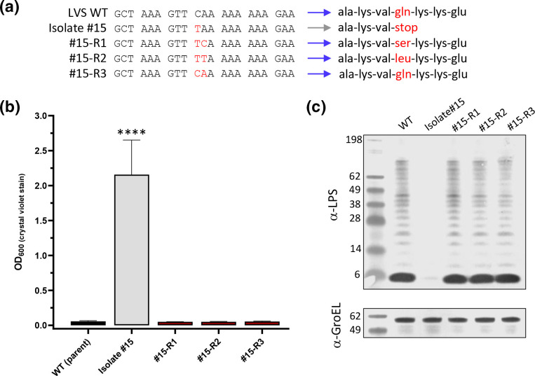
Natural reversion events within wbtJ can be detected in macrophage passaged isolate #15 samples. Heterogenous mixture of big and small colonies was observed in samples plated from macrophage assays with isolate #15 each time an experiment was conducted. Colonies that appeared larger than the parental isolate #15 were streak purified. (a) Sanger sequencing was performed of the wbtJ gene to determine the allele of the reverted isolate. Red font indicates a substituted nucleotide and the resulting codon compared to the LVS wild-type sequence for this gene. Blue and grey arrows indicate colony phenotype (b) The ability of the reverted isolates to form biofilm was assessed using crystal violet staining. For statistical analysis, a linear mixed effects model was used to compare each strain. ****p<0.0001. (c) The O-Ag was also assessed by western blotting performed with α-LPS (mAb FB11; top). α-GroEL (bottom) was included as a loading control.

