
Figure 2.
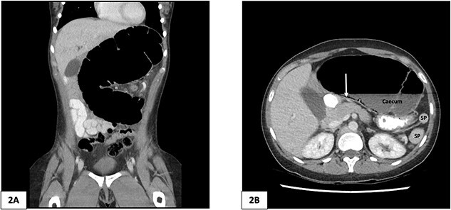
Computed tomography of the abdomen and pelvis of 25-year-old female patient. (A) Coronal slice demonstrating caecal volvulus with distended loop of colon. (B) Axial slice demonstrating 97 mm diameter distended caecum, multiple splenic fragments, arrow indicates pneumatosis coli, indicating colonic ischaemia. SP, spleen fragments.