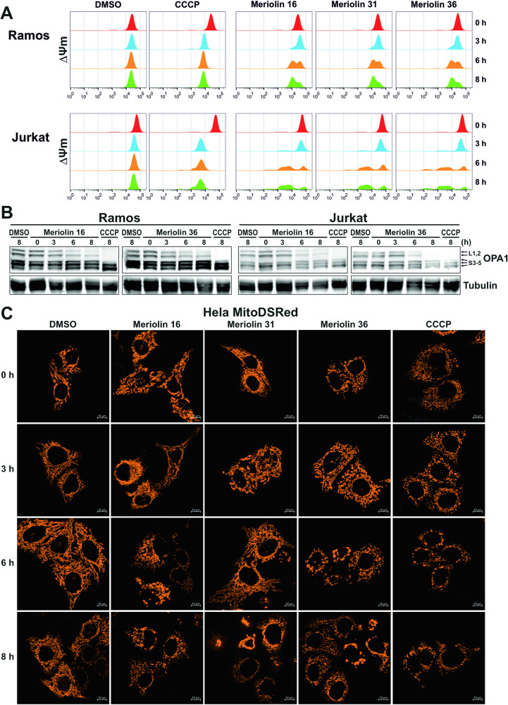
Fig. 6. Meriolin 16, 31, and 36 impair mitochondrial structure and function.
A Monitoring of the mitochondrial membrane potential (ΔΨm) of Ramos and Jurkat cells upon addition of 10 µM of meriolin 16, 31, or 36, 0.1% (v/v) DMSO (diluent control) or 10 µM CCCP (mitochondrial uncoupler, positive control) by flow-cytometric measurement of TMRE fluorescence. B The kinetics of the cleavage of the long isoforms (L1,2) of the dynamin-like GTPase OPA1 was determined by immunoblotting in Ramos (left panel) and Jurkat cells (right panel). Cells were treated as in (A) for the indicated time points. Immunoblotting for tubulin was used as a loading control. C Meriolin 16, 31, and 36 induce mitochondrial fragmentation (fission) in HeLa cells, stably expressing the fluorescent dye mito-DsRed targeted to the outer mitochondrial membrane. Cells were treated with DMSO (0.1% v/v), 1 µM of meriolin 16, 31, or 36, or 10 µM CCCP (positive control) for the indicated time points and mitochondrial morphology was assessed by microscopy (Apotome, Zeiss Axiovert). Shown are representative images.

