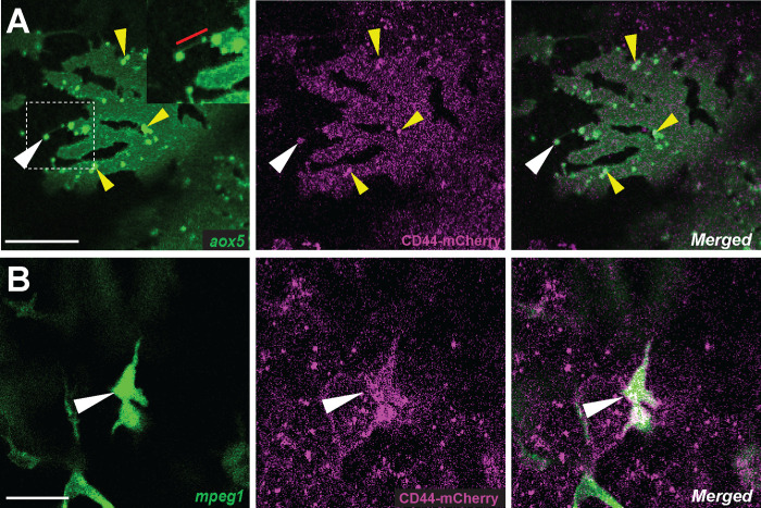
Figure 2. Localization of CD44a protein in xanthoblasts and metaphocytes.
(A) CD44-mCherry expression across the xanthoblasts cell membrane. CD44 is also notably present in the airineme vesicle (white arrowhead) and in the airineme blebs (yellow arrowheads). (B) CD44-mCherry expression in metaphocytes recognized by their amoeboid morphology, in contrast to the non-displayed dendritic population (white arrowheads) (Bowman et al., 2023). Scale bars represent 20μm (A, B).

