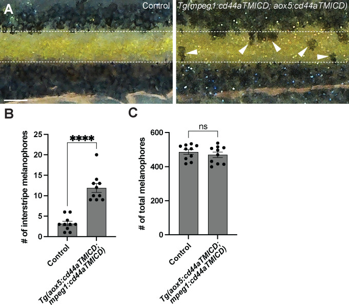
Figure 5. CD44-mediated airineme extension contributes to zebrafish pigment pattern formation.
(A) Unlike the control, melanophores failed to coalesce into stripe and remained in the interstripe zone (white arrowheads) at SSL11 (Parichy et al., 2009). The white dotted lines demarcate stripes and interstripe. (B) In embryos overexpressing cd44aTMICD simultaneously in both xanthophore-lineages and macrophage, the count of interstripe melanophores was significantly higher, (P<0.0001, 20 embryos in total). (C) However, the total number of melanophores was not differ in the experimental group as compared to the controls, (P=0.4525, 20 embryos in total). Statistical significance was assessed using a Student’s t test. Scale bars represent 200μm. Error bars indicate mean ± SEM.

